Compass:
Prostate Cancer Detection Needs Multi-View Context

Paul F. R. Wilson*1,6, Mohamed Harmanani*1,6, Zhuoxin Guo1,6, Obed K. Dzikunu2,6, Hannes Cash3, Adam Kinnaird4, Brian Wodlinger5, Purang Abolmaesumi†2, Parvin Mousavi†1,6
1Queen's University    2University of British Columbia    3Otto von Guericke University Magdeburg
4University of Alberta    5Exact Imaging    6Vector Institute
*Co-first authors    Co-senior authors

TL;DR - We introduce Compass, a study-level micro-ultrasound framework that jointly reasons over rotational prostate sweeps and biopsy frames. By conditioning mixed-evidence transformer aggregation on probe roll angle, Compass improves patient-level clinically significant prostate cancer detection.
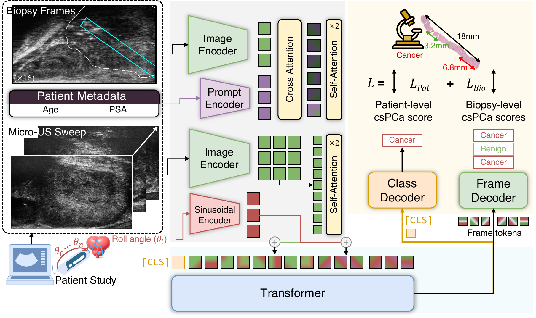
Compass architecture overview

Abstract

Artificial intelligence analysis of micro-ultrasound has shown promise for prostate cancer detection. However, most existing AI methods focus on the analysis of single images in isolation. By contrast, expert readers typically assess a full recorded video study, which provides three-dimensional context, to improve prostate cancer detection compared to single-frame analysis.

Inspired by this clinical workflow, Compass models a micro-ultrasound study as a stream of 2D images. Compass jointly integrates rotational sweep videos of the prostate with frames acquired at the moment of biopsy, and performs evidence aggregation across the study using a transformer conditioned on the probe's rotational angle. Trained and evaluated on a multi-center clinical trial dataset, Compass highlights the value of multi-view context for micro-ultrasound prostate cancer detection.

Method

Compass dual-branch architecture

Compass integrates two complementary sources of evidence: rotational micro-ultrasound sweeps that provide dense multi-view coverage of the gland, and biopsy core frames paired with clinical metadata. A frozen ProstNFound+ backbone encodes visual evidence, sinusoidal roll embeddings encode probe geometry, and a mixed-token transformer fuses biopsy and sweep tokens for patient-level and biopsy-level prediction.

Results

Patient and biopsy-level classification performance. Results are mean ±std over 5-fold cross-validation.
Method Patient-level Biopsy-level
AUROC B.Acc. Sen@60 AUROC B.Acc.
CLIP ViT-L/1467.9 ±7.971.3 ±5.357.9 ±18.062.6 ±11.462.9 ±8.0
Cinepro72.6 ±6.576.9 ±8.368.2 ±13.168.6 ±7.767.6 ±4.2
MedSAM74.6 ±5.876.8 ±5.343.1 ±24.461.7 ±4.361.6 ±3.1
ProstNFound+78.7 ±15.482.9 ±12.576.7 ±17.868.8 ±10.365.9 ±6.6
Cinepro-MIL76.3 ±13.976.1 ±11.273.3 ±19.2--
MedSAM-MIL75.7 ±13.776.4 ±10.373.3 ±19.2--
ProstNFound+-MIL77.3 ±14.578.1 ±9.482.5 ±15.3--
ViViT61.9 ±13.966.0 ±10.461.7 ±23.1--
3D-ResNet1871.1 ±6.072.3 ±4.673.2 ±11.9--
MicroSegNet+ViViT67.7 ±7.969.0 ±5.266.8 ±11.8--
ProstNFound++ViViT66.4 ±15.368.8 ±9.566.6 ±17.6--
PRI-MUS (Experts)78.5 ±11.178.3 ±6.983.8 ±15.873.1 ±8.971.9 ±7.5
Compass (ours)87.2 ±8.785.0 ±8.289.9 ±10.070.5 ±8.668.3 ±6.9

Compass achieves the strongest patient-level performance in this cohort. Its gains over frame-level, MIL, and video baselines suggest that sweep context and roll-aware cross-branch reasoning provide complementary information beyond isolated biopsy frames.

Ablation

Method Sweeps Angle PE Transformer AUROC Bal.Acc.
No angle embeddingyesnoyes82.775.1
No transformeryesyesno74.366.2
No sweepsnoyesyes78.870.6
Compass (ours)yesyesyes87.285.0

Removing the roll-angle embedding, transformer fusion, or sweep evidence degrades performance, indicating that Compass benefits from the combination of multi-view evidence, acquisition geometry, and cross-branch reasoning.

Clinical Evaluation

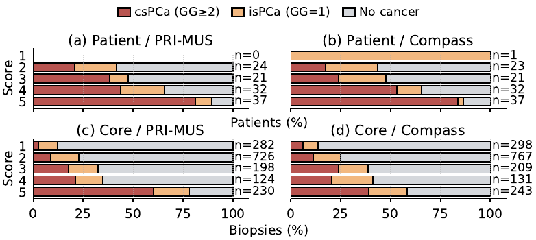
Outcome distributions across PRI-MUS and Compass score bins

Outcome distributions across score bins for PRI-MUS and Compass at the patient and core levels. Compass shows a clear patient-level risk gradient, while PRI-MUS remains stronger for localized core-level characterization, supporting Compass as a complementary study-level triage tool.

Qualitative Analysis

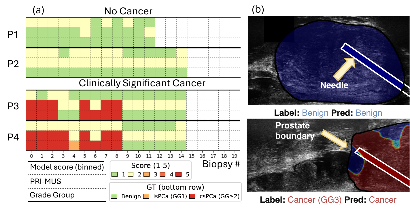
Compass risk scores and heatmaps across biopsy cores

Compass risk scores across biopsies within patients generally align with PRI-MUS risk scores and pathology findings. Post-hoc heatmaps visualize image-level cancer evidence conditioned on Compass embeddings.

BibTeX

@inproceedings{wilson2026compass,
  title   = {Compass: Prostate Cancer Detection Needs Multi-View Context},
  author  = {Wilson, Paul F. R. and Harmanani, Mohamed and Guo, Zhuoxin and Dzikunu, Obed K. and Cash, Hannes and Kinnaird, Adam and Wodlinger, Brian and Abolmaesumi, Purang and Mousavi, Parvin},
  booktitle = {International Conference on Medical Image Computing and Computer-Assisted Intervention (MICCAI)},
  year    = {2026},
}